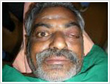
 Patients with Carotico Cavernous Fistula (CCF) present with a pulsatile swelling in the eye . The eye is usually red and swollen. It can happen suddenly and is often associated with a head injury. At times a CCF can be secondary to a spontaneous rupture of an abnormal artery into the vein. Rarely though, multiple small communications can develop between the blood vessels around the eye and the veins, leading to the same clinical symptoms. Treatment for CCF is always through the endovascular route. Surgery is limited to those situations where interventional radiology procedures cannot be performed.
Procedures Details
Presentation:
Patients with Carotico Cavernous Fistula (CCF) present with a pulsatile swelling in the eye . The eye is usually red and swollen. It can happen suddenly and is often associated with a head injury. At times a CCF can be secondary to a spontaneous rupture of an abnormal artery into the vein. Rarely though, multiple small communications can develop between the blood vessels around the eye and the veins, leading to the same clinical symptoms.
It is mandatory that such patients are evaluated at the earliest . The single most important investigation is angiography. Here a small tube is inserted into the blood vessel of the thigh under local anesthesia and navigated under image guidance to the blood vessels of the brain. A medicated dye (iodinated contrast) is then injected and multiple images taken.
These images are then analyzed not only to confirm the diagnosis but to also classify the type of CCF and to plan the treatment.
Treatment :
The aim of therapy is to stop the abnormal communication. The appropriate treatment would depend on the type of CCF.
In type I bed there is a large communication between the artery and the main, this would mean blocking the abnormal venous sac with either a detachable balloon or specially designed delicate platinum coils which are deployed through tubes taken from the blood vessel in the thigh ,like in angiography. In the other types Sometimes the feeding artery make be blocked with special glues.
The procedure is generally carried out under local anesthesia; however if coils or glue is the embolic agent of choice, the patient may require general anesthesia.
The patient may also be advised to compress the artery and vein in the neck intermittently since this has shown benefit in small fistulas.
Complications :
Though rare any Neuro-vascular interventional procedure carries the following risks.
- The procedure may fail and one may have to resort to surgery.
- A normal vessel may get blocked and the patient may develop a stroke due to lack of supply to that region.
- The area may get worse due to a blood clot passing into normal vessels and may even result in death
- The vessel being treated may rupture and can result in death.
- Drugs including the dye used for angiography can produce an allergic reaction which can rarely be severe and dangerous to life.
- Patient may develop bleeding from the site of puncture for angiography in the leg or arm; this can be stopped by compression for a few minutes.
Appointment :
Information Contact : dr.mathewcherian@gmail.com
Phone : +91 9600900373,+91 422 2628627
Case Study
Type D CCF